
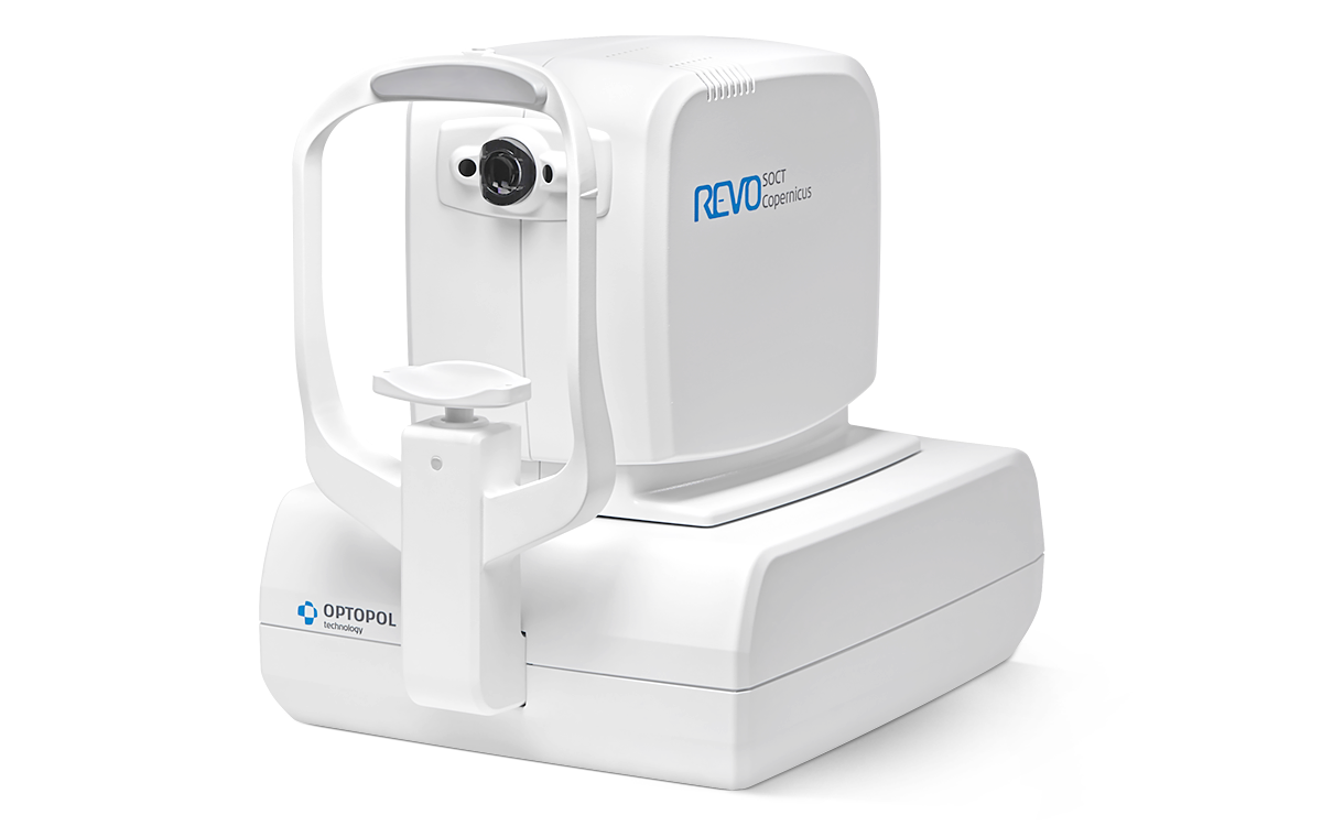

Optopol SOCT Copernicus REVO
 Or please call FAISAL: 079 242 2817 or AHMED: 082 414 1472
SOCT Copernicus REVO offers all the newest standards available in Spectral OCT technology.
![]() Click here to view the pdf catalogue
Click here to view the pdf catalogue
Our supreme experience in Spectral Domain OCT technology allows us to provide you with the modern OCT that offers remarkable simplicity of operation. The new SOCT Copernicus REVO will meet the daily demands of any modern practice.
OCT made simple as never before
Position the patient and press the START button to acquire examinations of both eyes.
The SOCT Copernicus REVO, using vocal messages, guides the patient through the process increasing comfort and reducing patient chair time.
Creating customised scanning protocols of different diagnostic scenarios will speed up workflow.
A perfect fit for every practice
Small system footprint, various operator and patient positions and connection by a single cable allows the installation of SOCT Copernicus REVO into the smallest of examination room spaces. Revo’s variety of examination and analysis tools enables it to effortlessly function as a screening or advanced diagnostic device.
High quality of OCT image
The noise reduction technology provides the finest details proven to be important for early disease detection.
RETINA
A single 3D macula scan performs both Retina and Glaucoma analysis.
The software automatically recognises 8 retinal layers which assists with a precise diagnosis and the mapping of any changes in the patient’s condition.
ANGIOGRAPHY SOCT
This non-invasive dye free technique allows the visualization of the microvasculature of the retina. Both blood flow and structural visualization will give additional information in the diagnosis of many retinal diseases. Angiography scan allows assessment of the structural vasculature of the macula, periphery or the optic disc. Extremely short scanning time 1.6 second in standard resolution or within ~3 seconds in
high resolution.
Now Angiography OCT can become a routine diagnosis in your practice.
ANGIOGRAPHY MOSAIC
The Angiography mosaic delivers high-detailed images over large field of the retina. Available modes allow to see predefined region of the retina in a convenient way. Manual mode allows to scan desired region. Analysis tools allow to see vascular layers, enface or thickness maps.
WIDEFIELD SCAN
12x12 mm Widefield Central scan is perfect for fast and precise screening of the patient’s retina.
GLAUCOMA
Comprehensive glaucoma analysis tools for quantification of Nerve Fiber Layer, Ganglion layer Optic Nerve Head with DDLS allows for precise diagnosis and the monitoring of glaucoma over time.
Asymmetry Analysis of Ganglion layers between hemispheres and between eyes allow the identification and detection of glaucoma in its early stages and in nontypical patients.
FOLLOW UP
Revo’s standard high density scanning capability and blood vessel structure recognition enable a precise alignment of past and current scans Operator can analyze changes in morphology, quantified progression maps and evaluate the progression trends Progression Morphology Progression Quantification
ANTERIOR
For a standard anterior examination, no additional lens is required. This allows the examiner to quickly complete the scanning procedure.
Additional adapter provided with the device increases range of clinical application in Anterior chamber observation.
BIOMETRY OCT
B-OCT® Innovative method of using the posterior OCT device to measure ocular structure along eye axis. OCT Biometry provides complete set of Biometry parameters: Axial Length AL, Central Cornea Thickness CCT, Anterior Chamber Depth ACD, Lens Thickness LT.
VISUALLY VERIFY YOUR MEASUREMENT
All measurement calipers are shown on all boundaries of OCT image provided by REVO. Now, you can visually verify, identify and if needed correct which structure of the eye has been measured.
